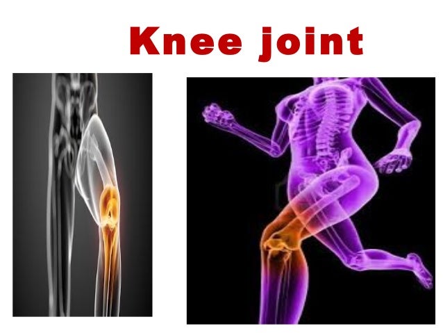

Posterior suprapatellar (prefemoral or supratrochlear) fat padĪnterior suprapatellar (quadriceps) fat padĬonnect the anterior limbs of the two menisciĪnterior (Humphrey) and posterior (Wrisberg) meniscofemoral ligaments: The cruciate ligament and popliteus tendon are extrasynovial but intracapsularĬommunicates with the suprapatellar bursa Joint capsule is lined by synovial membrane, however, the attachment of the synovial membrane does not coincide with the capsular attachments because of the intra-articular structures One communicating with suprapatellar bursa Laterally the capsule is not attached to the tibia but is prolonged down over the popliteus tendon Posteriorly to the ridge between the two condyles at the lower end of the groove for the PCL On the lateral condyle it encloses a pit and groove for the popliteus tendonĪttached around the margins of the tibial plateau except in two places Posteriorly attached to the intercondylar ridge at the lower limit of the popliteal surface The medial meniscus is attached to the medial collateral ligament and the lateral meniscus is attached to the popliteus tendonĪttached to the femur and tibia via the coronary ligamentsĪdheres below the epiphyseal line down to the articular margin except in two places With extension, the contact area lessens and moves distallyįibrocartilaginous, C-shaped in appearance and triangular in cross-section On flexion, more parts of the bony surface are exposed to articulation (four below, odd facet) and are more proximal on the patella hamstring group: A group of three muscles found in the posterior region of the thigh, responsible for flexing of the lower leg at the knee. Medial, lateral and odd facet on the posterior surface of the patella articulate with the medial and lateral condyles of the femur Saddle joint between the patella and femoral condyles: Laterally: between a wide and flat femoral condyle and a circular tibial articular surface which overhangs the shaft posterolaterally Medially: between a narrow and curved femoral condyle, and an oval tibial articular surface with a long anteroposterior length

There are medial and lateral articular facets on the tibial plateau and medial and lateral femoral condyles on the distal femur with are convex and circular shaped. There are two condylar joints between the femur and tibia (tibiofemoral). Movement: flexion to 150°, extension to 5-10° hyperextension rotation whilst in the flexed position to 10° actively and 60° passively Nerve supply: branches from the femoral, tibial, common peroneal, and obturator nerves Location: two condylar joints between femur and tibia saddle joint between patella and femurīlood supply: main supply are the genicular branches of the popliteal artery


 0 kommentar(er)
